
CBCT imaging and computer-based planning are the single biggest improvement in implant dentistry over the past 20 years. CBCT shows the precise 3D anatomy of your jaw, sinus, and nerves — measured down to the millimeter — and 3D virtual planning software turns those measurements into a case designed before a single instrument is picked up. Every implant case at this practice is planned this way.
Book Now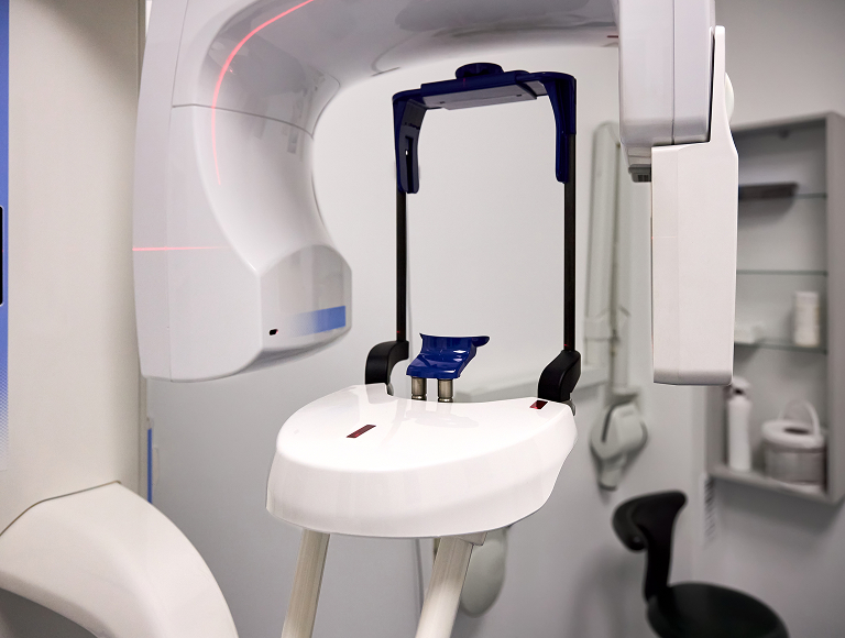
Standard dental X-rays show a flat, 2D shadow. CBCT reveals the full picture needed for safe, precise implant placement.
Bone height and width at every potential implant site, measured to the millimeter
Bone density in different jaw regions, which affects implant primary stability
Inferior alveolar nerve location in the lower jaw
Maxillary sinus dimensions and floor position in the upper jaw
Adjacent tooth roots and their angulation
Anatomical variations that affect surgical access (sinus septa, ridge undercuts, narrow alveolar processes)
Your CBCT data is imported into 3D virtual planning software, where the implant is placed digitally at its optimal position, depth, and angulation — checked against nerve position, sinus floor, adjacent roots, and the final tooth before surgery day. That plan is what guides placement in the chair, so the surgery follows a design that was already verified in three dimensions.
Book NowA CBCT for implant planning delivers roughly the same radiation as 3–5 days of natural background radiation, and about 1/10th the dose of a routine medical CT. Lead aprons, thyroid collars, and dose-optimized protocols are used to minimize exposure further.
Book Now
Schedule a consultation at Tuscaloosa Dentures and Implants today.
Free Implant Consultation
Your consultation and 3D CBCT scan are completely free — so you know exactly where you stand before deciding anything.